Over the past three decades, many studies have reported a high success rate for rehabilitation with zygomatic implants. (Image: Anna Jurkovska/Shutterstock)
The presence of sufficient bone volume is one of the most important criteria for successful osseointegration of implants,1 wherefore restoration of atrophied edentulous maxillae poses a great dilemma for the surgeon and restorative dentist. Sinus bone grafting to build new bone for implant anchorage in atrophied jaws entails multiple surgical interventions and has varying implant success rates, high potential for donor site morbidity and increased surgical costs.2, 3 A major breakthrough came when Brånemark first used custom-designed, longer implants inserted into the zygomatic bone in support of a craniofacial prosthesis in the 1980s.4 When used in the treatment of maxillary atrophy,5, 6 zygomatic implants present a graftless alternative.
Zygomatic implant concept
The zygomatic implant design and placement protocols have been extensively described.6–17 In short, the implant, ranging from 30.0 mm to 62.5 mm, is introduced into the second premolar area, traversing the maxillary sinus, and is anchored in the zygomatic bone. In addition to two zygomatic implants, two to four conventional implants are required in the anterior maxilla to support the prosthesis.18, 19 Zygomatic implants have shown good clinical success rates in clinical studies, most often close to 100% success in follow-up periods of up to five years.20–23 Sinuscopy performed in patients with zygomatic implants has shown absence of infection or inflammation in the surrounding mucosa.24 Furthermore, placement of multiple (two to four) zygomatic implants in the same zygomatic bone has been reported to be a clinically successful treatment and to have similar complications to those experienced with the original technique.25–29
Immediate function, a well-documented concept associated with the immediate loading of implants upon insertion,30–33 has shown high success rates, provided that there is high initial stability. Histological analysis of the zygomatic bone shows a regular trabecular structure and a larger compact bone density (up to 98%).34 Owing to the high osseous density of zygomatic bone34 and the high clinical survival rates associated with zygomatic implants,20–23 this tissue–implant interface is particularly suitable for immediate function.
Dynamic navigation
Three-D implant planning and mapping are two important steps in implant rehabilitation,35, 36 in the sense that they represent a preliminary stage to actual surgery. Misplaced implants can create difficult aesthetics and functional and biological problems and may result in implant loss.37–39 There are three ways to transfer a planned implant’s position into the actual patient’s jawbone: (a) mental navigation, called freehand navigation; (b) static navigation using surgical templates;40 and (c) dynamic navigation.41, 42
The freehand approach is totally dependent on the surgeon’s experience, skills and mindset during treatment and creates the highest deviations compared with the other approaches.36 The use of surgical templates provides a higher accuracy compared with freehand surgery, but has several limitations, such as the inability to modify the plan once the surgical template has been manufactured. Surgical templates require longer drills, which can make their use difficult in patients with limitations in terms of mouth opening. Other concerns are irrigation issues and incompatibility with advanced surgical protocols. Dynamic navigation is, at present, the most effective way to transfer the planned implant’s position to the actual patient, as it guides the surgeon’s motions using real-time feedback. It is especially useful for reducing flapped procedures and offers the advantage of improved soft-tissue healing and patient comfort and reduction in bone resorption. Dynamic navigation allows modification planning at any time, even during treatment, and can be used in cases with limited mouth opening or in combination with osseodensification drills.
StealthStation S8 system
The StealthStation S8 surgical navigation system (Medronic) enables precise tracking of the location of technical instruments throughout a surgical procedure. The system introduces a combination of hardware, software, tracking algorithms, image data merging and specialised instruments to help guide the surgeon during surgical procedures.43, 44 The StealthStation S8 system is intended as an aid for precisely locating anatomical structures in either open or percutaneous procedures. The system is indicated for any medical condition in which the use of stereotactic surgery may be appropriate and where reference to a rigid anatomical structure, such as the skull, a long bone or a vertebra, can be identified relative to a tomography- or magnetic resonance-based model, fluoroscopic images or digitized landmarks of the anatomy.43, 44
For the software to display the instrument’s location in relation to the patient’s images, the software must create a map between the points on the patient and the points on the images. This process is called registration. Upon completion of registration, whenever the operator touches a point on the patient using a special tracked instrument, the computer will use the map to identify the corresponding point on the images. This identification is called navigation. A navigated point is identified on the system screen in various patient image planes and other anatomical renderings.
The surgical navigation system offers both optical and electromagnetic tracking capabilities, integration with external devices like microscopes and ultrasonic devices, a broad array of instrument offerings, and core software applications for neurosurgery, spine procedures and maxillofacial surgery.43, 44 During navigation, the system identifies the location of the tip and the trajectory of the tracked instrument in images and models that the user has selected for viewing. The surgeon may also create and store one or more surgical plane trajectories prior to surgery and simulate the progression along these trajectories. In surgery, the software can show the actual position at the tip of the instrument and its trajectory, relating them to the preoperative plane.43, 44
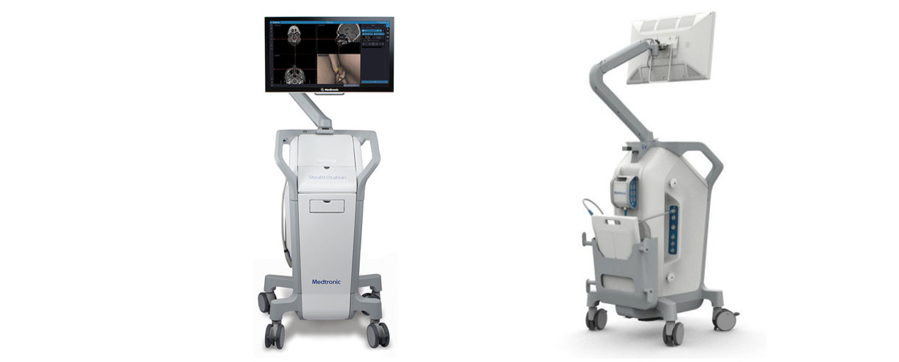
Fig. 1: StealthStation S8 system (Medtronic) equipment.
To maintain accuracy, the StealthStation S8 system uses dynamic referencing to constantly track the position of the anatomy during registration and navigation. Two devices are required for dynamic referencing: a patient frame of reference and a locator. The patient’s frame of reference is rigidly positioned in relation to anatomy. The locator, which is a camera for optical tracking or a transmitter and an instrument interface for electromagnetic tracking, finds the patient’s frame of reference and reports the position of the frame to the navigation software.
As the frame of reference remains in a rigid and fixed position in relation to the anatomy, any movement of the anatomy or of the locator results in corresponding movement of the frame of reference in the locator’s field of view. This allows the locator to detect any movement of the anatomy by identifying the position of the frame of reference, which moves simultaneously with the anatomy. The system can then display the instrument or implant location relative to the patient’s frame of reference while maintaining accurate navigation. Without dynamic referencing, any movement of the locator after registration would be invalid, as the position of the frame of reference would be changed in the navigation field. Dynamic referencing allows the flexibility to reposition the locator at any time. The system consists of a platform, clinical software, surgical instruments and a reference system (including patient and instrument trackers; Fig. 1). Three-D images of the patient can be displayed by software from various perspectives (axial, sagittal, coronal and oblique planes).43, 44
Clinical case
A 56-year-old male patient attended the oral-maxillofacial surgery consultation at Clitrofa medical centre in Trofa in Portugal to perform an implant-supported rehabilitation of the upper jaw. The clinical evaluation revealed a partially edentulous jaw with the presence of teeth #11, 12, 13, 21 and 24, which supported a removable prosthesis (Fig. 2).
To complete the preoperative evaluation, a high-definition CT scan was performed, which revealed an extremely resorbed maxilla in the posterior. Placement of two zygomatic implants in the posterior and four conventional implants in the anterior sector of the maxilla was indicated (Fig. 3). The CT scan was uploaded to StealthStation S8, and matching between the patient’s actual anatomy and imaging was performed. The flat emitter was placed below the patient’s head to eliminate obstructions for pinless and surgical workflows (Fig. 4).
Figs. 2a–c: Pre-op evaluation: dental occlusion (a); dental panoramic tomograph (b); and appearance of the contour of the lips and orbicularis oris muscle of the mouth (c).
Figs. 3a–c: Initial CT scan with cross-sectional (a), coronal (b) and sagittal (c) views.
Fig. 4: StealthStation S8 matches the CT imaging with the patient’s actual anatomy.
Fig. 5: The navigation instrument.
Figs. 6a & b: Placement of the first-quadrant zygomatic implant with the help of the navigation instrument.
Figs. 7a & b: Placement of the second-quadrant zygomatic implant with the help of the navigation instrument.
Fig. 8: Intra-op aspect of implant placement.
Fig. 9: Three-D intra-op images provided by the navigation system.
Figs. 10a–c: Final CT scan with cross-sectional (a), coronal (b) and sagittal (c) views.
After elevation of a full-thickness flap with bilateral identification of the infraorbital nerves, an osteotomy was performed to create a bone window to access the interior of the maxillary sinus. The navigation instrument most suitable for this type of surgery was chosen for its flexibility, thickness and length (Fig. 5). The images displayed on the monitor are in real time intra-operatively, allowing the alteration and verification of the osteotomy in the space planes. Confirmation of existing bone availability, maintenance of the integrity of the relevant anatomical structures and placement of the zygomatic implant in the ideal position for each clinical case are ensured. Zygomatic implants are placed according to this checklist (Figs. 6a–7b). The 3D positioning of the zygomatic implants allowed excellent primary stability as well as adequate positioning for prosthetic restoration (Fig. 8).
The images provided intra-operatively and in real time are of great definition and highly informative. The system also allows the introduction of a colour code to establish safety limits with respect to the length and diameter of the zygomatic implants. Navigation can also be used for other conventional implants (Fig. 9). After completion of the surgery, a new high-definition CT scan was performed to check the final positions of the two zygomatic implants placed in the posterior of the maxilla and the four conventional implants placed in the anterior sector of the maxilla (Fig. 10).
Conclusion
The StealthStation navigation system is an intra-operative advantage in the placement of the zygomatic implant and can form part of surgical protocols in its current form. However, there are some aspects that should be improved, namely the incorporation of a virtual library with the dimensions of the available zygomatic implants and the adaptation of the navigation system to the contra-angle handpiece used in this type of surgery.
HELSINKI, Finland: Planmeca has introduced two dental chair units designed to give clinics more choice in dental chair unit configuration, treatment room ...
GOTHENBURG, Sweden: The 2026 Implant Solutions World Summit opened yesterday at the Swedish Exhibition and Congress Centre, bringing together more than ...
Reliable reprocessing of handpieces and turbines remains a significant challenge for many practices, particularly amid strict regulatory requirements and ...
Keystone Dental, a global medical technology company, has launched the Keystone Dental Campus, a new online education platform for dental professionals. The...
As pathological tooth wear becomes a more frequent concern in clinical practice, direct composite restorations are increasingly relevant as a minimally ...
CAD/CAM technology continues to influence how dental practices and laboratories plan, design and deliver restorative treatment, and dental teams need to ...
As digital technologies become more embedded in dental diagnostics, treatment planning and restorative workflows, new questions are emerging about how ...
Alliedstar, a provider of advanced scanning and CAD/CAM solutions, has launched the Alliedstar Academy, a new online education platform designed to help ...

 International / International
 Brazil / Brasil
 Canada / Canada
 Latin America / Latinoamérica
 USA / USA
 Austria / Österreich
 Bosnia and Herzegovina / Босна и Херцеговина
 Bulgaria / България
 Croatia / Hrvatska
 Czech Republic & Slovakia / Česká republika & Slovensko
 France / France
 Germany / Deutschland
 Greece / ΕΛΛΑΔΑ
 Hungary / Hungary
 Italy / Italia
 Netherlands / Nederland
 Poland / Polska
 Portugal / Portugal
 Romania & Moldova / România & Moldova
 Slovenia / Slovenija
 Serbia & Montenegro / Србија и Црна Гора
 Spain / España
 Switzerland / Schweiz
 Turkey / Türkiye
 UK & Ireland / UK & Ireland
 China / 中国
 India / भारत गणराज्य
 Pakistan / Pākistān
 Vietnam / Việt Nam
 ASEAN / ASEAN
 Israel / מְדִינַת יִשְׂרָאֵל
 Algeria, Morocco & Tunisia / الجزائر والمغرب وتونس
 Middle East / Middle East

 Dr. Fernando FranchLive webinar
 Dr. Nicolas OuelletRegister now1CELive webinar

 Dr. Nisha D’Silva BDS, MSD, PhD, Dr. Kıvanç Bektaş-KayhanRegister now1CELive webinar
 Federico ZunicaRegister now1CE


; dental panoramic tomograph (b); and appearance of the contour of the lips and orbicularis oris muscle of the mouth (c).")
, coronal (b) and sagittal (c) views.")


, coronal (b) and sagittal (c) views.")
; dental panoramic tomograph (b); and appearance of the contour of the lips and orbicularis oris muscle of the mouth (c).")
, coronal (b) and sagittal (c) views.")


, coronal (b) and sagittal (c) views.")


To post a reply please login or register