A recent study has found that reducing the patient radiation to 20% of the manufacturer-recommended protocol does not compromise the CBCT image quality. (Image: Massimo Cattaneo/Shutterstock)
MALMÖ, Sweden: Radiographic diagnostics are widely used in healthcare as they provide diagnostically important information that can help improve treatment outcomes. For example, a CBCT scanning protocol is a valuable examination tool in oral and maxillofacial radiology and is readily available in dental offices because of its ease of use. However, a CBCT scan produces a relatively high radiation dose compared with other scanning protocols, and new research has shown that its effectiveness remains unchanged after reducing the radiation dose to one-fifth of the manufacturer-recommended level.
To get a better look at an injury site, a surgeon often refers a patient for CBCT scans. For example, a CBCT scan can be a valuable tool for diagnosis and treatment planning for patients with temporomandibular disorders as it helps clinicians to perform assessments of bone changes with high diagnostic accuracy.
“With the CBCT, we take pictures of the mouth and jaw from three angles—from the front, from below, and from the side; this is in order to be able to answer the question that comes with the referral,” Dr Kristina Hellén-Halme, senior lecturer in the Faculty of Odontology at Malmö University, said in a press release. She went on to add that a radiographic examination is not a risk in itself but that it should not be taken without a valid reason.
Since the use of CBCT scans in dentistry is growing, a recent study sought to evaluate whether the high dose of patient radiation that is often used in CBCT imaging is really necessary to provide acceptable image quality.
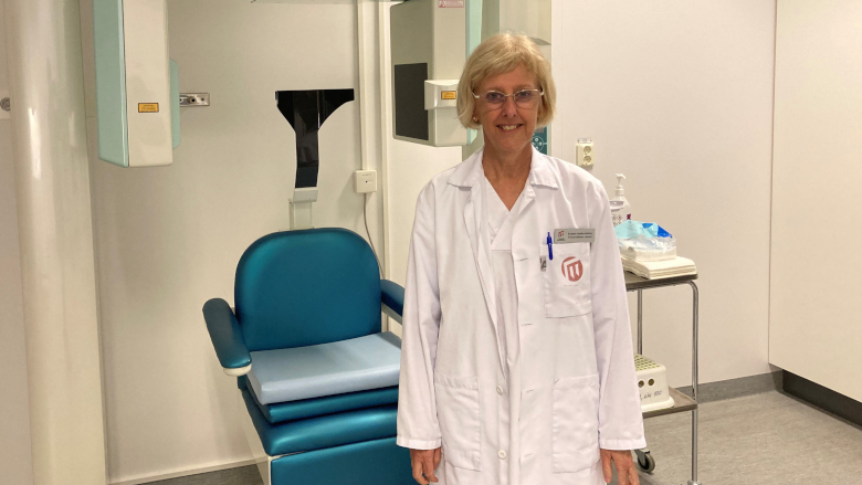
Dr Kristina Hellén-Halme (Image: Hanna Svederborn)
“Many different scanning protocols are available, and sometimes the urge from clinicians to have more or less noise-free images can increase the radiation dose to patients without adding more information to the specific aim of the investigation,” the researchers explained.
In the study, 34 adult patients referred for CBCT imaging of the temporomandibular joint underwent two examinations with two scanning protocols: a manufacturer-recommended protocol and a low-dose protocol where the tube current was reduced to 20% of the recommended protocol. The researchers noted that manufacturer-recommended exposure settings for the CBCT scanners vary widely and may result in varying amounts of radiation doses.
After evaluating the visibility of temporomandibular joint anatomic structures and image quality, the radiologists who were tasked to analyse the images concluded that both the low and high dose images yielded diagnostically comparable results. Therefore, the findings suggest that high image quality is not necessary for all clinical situations and that dental professionals should reduce the recommended patient radiation dose provided that it does not affect diagnostic outcomes. They noted that this is especially important when treating younger patients who are at a higher risk from radiographic exposure than adults owing to the radiosensitivity of their developing tissues.
AARHUS, Denmark: As the implementation of artificial intelligence (AI) in the dental industry continues to accelerate, there appears to be a growing ...
UMEÅ, Sweden: As post-pandemic research continues to examine how different public health responses shaped oral healthcare access and outcomes, a new study ...
OSLO, Norway: Researchers have recently investigated attitudes and activities among dental healthcare professionals working in the public dental service ...
GOTHENBURG, Sweden: Older age is a major predictor of various health issues, including cognitive and cardiovascular disease, hearing loss and progressive ...
COPENHAGEN, Denmark: In a recent study, researchers examined the dentate status and the frequency of preventive dental visits of Danish adults over a period...
COPENHAGEN, Denmark: In Denmark, dental care regulations state that orthodontic treatment should ideally be led by orthodontists, but adult aligner ...
STOCKHOLM, Sweden: Ten years ago, Sweden did away with the recommendation of administering antibiotics prophylactically for patients deemed at risk of ...
These days, there is a growing emphasis on quality over quantity, that is, preferring an object or service that is worthwhile rather than a quick and easy ...
OSLO, Norway: The widespread availability of vaccines in developed nations has significantly changed the risk of dentists contracting SARS-CoV-2 in a ...
Education
Live webinar Monday, 13. July 2026 17:30 CET (Oslo)
HELSINKI, Finland: Planmeca has introduced two dental chair units designed to give clinics more choice in dental chair unit configuration, treatment room ...
GOTHENBURG, Sweden: The 2026 Implant Solutions World Summit opened yesterday at the Swedish Exhibition and Congress Centre, bringing together more than ...
Reliable reprocessing of handpieces and turbines remains a significant challenge for many practices, particularly amid strict regulatory requirements and ...

 International / International
 Brazil / Brasil
 Canada / Canada
 Latin America / Latinoamérica
 USA / USA
 Austria / Österreich
 Bosnia and Herzegovina / Босна и Херцеговина
 Bulgaria / България
 Croatia / Hrvatska
 Czech Republic & Slovakia / Česká republika & Slovensko
 France / France
 Germany / Deutschland
 Greece / ΕΛΛΑΔΑ
 Hungary / Hungary
 Italy / Italia
 Netherlands / Nederland
 Poland / Polska
 Portugal / Portugal
 Romania & Moldova / România & Moldova
 Slovenia / Slovenija
 Serbia & Montenegro / Србија и Црна Гора
 Spain / España
 Switzerland / Schweiz
 Turkey / Türkiye
 UK & Ireland / UK & Ireland
 China / 中国
 India / भारत गणराज्य
 Pakistan / Pākistān
 Vietnam / Việt Nam
 ASEAN / ASEAN
 Israel / מְדִינַת יִשְׂרָאֵל
 Algeria, Morocco & Tunisia / الجزائر والمغرب وتونس
 Middle East / Middle East

 Dr. Fernando FranchLive webinar
 Dr. Nicolas OuelletRegister now1CELive webinar

 Dr. Nisha D’Silva BDS, MSD, PhD, Dr. Kıvanç Bektaş-KayhanRegister now1CELive webinar
 Federico ZunicaRegister now1CE


To post a reply please login or register